
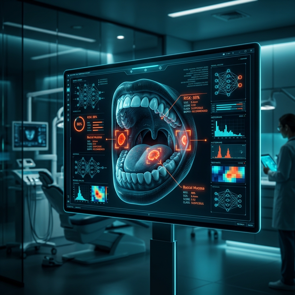
1. Study Objective & Overview
A 2026 systematic review published in Brazilian Dental Science evaluated artificial intelligence technologies applied to early oral cancer detection. The review focused on Oral Squamous Cell Carcinoma (OSCC) and Oral Potentially Malignant Disorders (OPMDs), investigating the clinical impact of computer vision and machine learning models in screening workflows.
Delayed diagnosis and limited access to specialist oral pathologists in remote areas remain significant clinical challenges, driving research into automated image analysis for point-of-care screening.
2. Results Reported in the Review
The review synthesized findings across studies evaluating deep learning models trained on clinical intraoral photographs, autofluorescence images, and histopathological slide data:
- Diagnostic Accuracy Generally >80%: Reported diagnostic accuracy across the reviewed literature was generally above 80% (ranging overall from 72.1% to 98.6%, with top hybrid and Transformer-based models reaching 85% to 99.1% depending on architecture and image modality).
- Multi-Modal Feature Extraction: Deep learning algorithms analyzed subtle visual variation in color, tissue margins, and mucosal texture.
- Point-of-Care Application: Reviewed studies highlighted mobile-compatible algorithms designed to assist healthcare personnel in conducting initial screening and triage in low-resource settings.
3. Discussion & Clinical Conclusion
The authors noted that while AI image classification demonstrates strong diagnostic performance, real-world deployment faces challenges regarding dataset heterogeneity, image quality standardization, and model generalization across diverse populations.
AI algorithms serve as valuable decision-support and triage tools. However, image-based AI cannot replace histopathological evaluation; definitive diagnosis requires clinical examination and scalpel tissue biopsy.
4. Synthesis of Key Takeaways
Mobile AI technologies are being developed as point-of-care triage tools to assist healthcare workers in screening oral mucosal lesions in regions with limited specialist access.
The review highlights how computer vision models extract visual image features from clinical, autofluorescence, and histological datasets to assist in lesion classification.
AI provides decision-support during initial triage (>80% accuracy). However, tissue biopsy and histopathology remain mandatory for definitive diagnosis.
Brazilian Dental Science, 2026. "AI in early oral cancer detection: a systematic review of technologies and clinical impact."